

💡 AI Summary & Key Takeaways
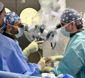
– Surgical operating microscopes provide high-definition magnification and coaxial illumination.
– Microdiscectomy selectively removes the herniated fragment while sparing the healthy disc annulus.
– Exceptional >95% success rate in providing immediate relief from radicular sciatic pain.
– Microdiscectomy selectively removes the herniated fragment while sparing the healthy disc annulus.
– Exceptional >95% success rate in providing immediate relief from radicular sciatic pain.
The Gold Standard in Spinal Decompression
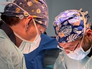
Microscopic Spine Surgery (Microdiscectomy) remains the international benchmark for herniated disc surgery. Employing multi-axis Carl Zeiss/Leica surgical microscopes provides up to 20x magnification, allowing unmatched distinction between neural tissues, herniated fragments, and epidural veins.
Targeted Fragmentectomy Technique
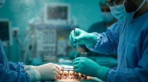
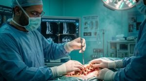
Through a 2-cm mini-incision, the surgeon executes a targeted sequestrectomy, preserving the spinal architecture and facet joints to maintain segmental biomechanical integrity.